
Translating molecular pathology into cancer diagnostics.
Pathology underpins 99 per cent of all management decision for cancer patients. As the understanding of cancer as a genetic disease has evolved, so too have approaches to its diagnosis, classification, and treatment stratification for patients.
Led by Professor Stephen Fox, the Translational and Diagnostic Pathology lab uses cutting edge molecular pathology to identify and develop new biomarkers for cancer diagnosis, prognosis and treatment to significantly improve patient outcomes.
The lab identifies biomarkers revealing the unique molecular characteristics of an individual’s tumour, forming the foundation of precision medicine. This enables them to make accurate diagnoses and match specific genomic changes with targeted therapies, ensuring selection of patients most likely to benefit from particular drugs. Equally important, their approach spares biomarker-negative patients from unnecessary toxicity by avoiding ineffective treatments.
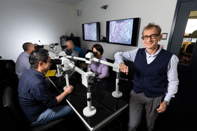
The Translational and Diagnostic Pathology Lab is based in the Peter MacCallum Cancer Centre.
Our research
-
The ductal carcinoma in situ (DCIS) project aims to understand the biology of this treatment dilemma—abnormal cells that can potentially develop into invasive, metastatic breast cancer. Risk of progression is hard to determine, leading to many patients being over-treated, and others not receiving sufficient treatment to prevent invasive recurrence.
The project aims to identify biomarkers to predict which patients will need aggressive treatment, and which can avoid toxic and costly side-effects from hormonal therapies and radiotherapy.
The lab use multiple conventional and novel techniques — such as spatial transciptomics and single cell methods — to identify potential biomarkers during the pivotol transition from in situ to invasive breast carcinoma.
Other technologies being explored are sheet microscopy for 3D rendering of tumour architecture and phenotype, and application of machine learning/artificial intelligence to DCIS digital pathology.
-
This program aims to translate new genomic technologies, assays, and tools into clinical practice to improve test efficiency, accuracy, turnaround times, and reporting for cancer patients. It involves developing and validating assays for early-phase clinical trials.
Key areas include using whole genome sequencing/transcriptomics to resolve diagnostic dilemmas, leveraging circulating tumour DNA for therapeutic decision-making when biopsies are unavailable, and monitoring neoadjuvant therapy response through circulating tumour cell profiling in breast cancer.
Priorities include comprehensive fusion assays, low-cost cancer gene panels, and evaluating long-read sequencing for germline and central nervous system tumours. Implementation studies will explore delivering complex genomic test information through multidisciplinary meetings. Workforce development and education underpinning clinical cancer genomics adoption are also critical components of this program.
Digital pathology
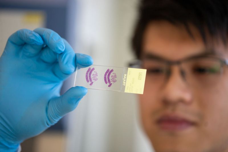
The pilot digital pathology program uses whole slide imaging (scanning and digitising tissue slides at high resolution), which enables pathologists to examine the details of tissue digitally, instead of at a microscope.
The program will enable:
- second opinion diagnoses
- automated validated quantitation of biomarkers used in clinical practice (as opposed to manual), and
- development of algorithms for tumour identification and quantitation for subsequent molecular profiling.
Latest News
Contact and more information
Professor Stephen Fox
Director of Pathology, Peter MacCallum Cancer Centre
stephen.fox@petermac.org